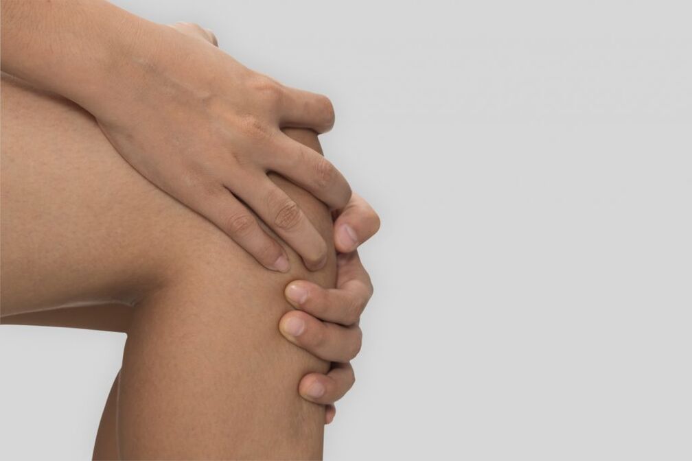
The knee joint experiences regular stress. Running and jumping, walking and climbing stairs, or just standing, all affect the condition of the cartilage tissue of the knee. If the balance of the cartilage is disturbed, the development of arthrosis - gonarthrosis - of the knee joint begins.
Gonarthrosis is a deforming arthrosis of the knee joint, which is accompanied by chronic damage to the surface of the hyaline cartilage and bones - the femur and the tibia. Symptoms of knee joint disease are pain that worsens with movement. Due to the accumulation of fluid in it, movement is limited. Later, there is a limitation of the movement of the knee due to a violation of the foot support. The diagnosis of the pathology is based on the collection of the patient's anamnesis and complaints, visual examination of the knee joint and hardware examination. Such age-related disorders of the locomotor system appear in almost everyone in old age.
general information
Gonarthrosis (from the Latin genus articulatio - knee joint) is the most common arthrosis, which is a degenerative-dystrophic progressive, non-inflammatory change in the cartilage of the knee joint. Women and the elderly usually suffer from gonarthrosis. But after injuries acquired during intense sports, gonarthrosis occurs even in young people.
The cause of arthrosis is a change in the structure of the cartilage within the joint, not the deposition of salts there. In the case of gonarthrosis, salt deposits form where the tendons are connected to the ligamentous apparatus, but they cannot cause the symptoms of pain. First, cracks appear in the cartilage, reducing the thickness in some areas. Gradually, the load is redistributed, the joint begins to come into contact with the bones, accelerating the painful process. As a result, the following changes occur in the knee joint:
- thinning of the knee cartilage until it disappears;
- changes in the composition and amount of synovial fluid;
- damage to the bones of the knee due to friction;
- appearance of osteophytes;
- stiffness due to compression of the joint capsule;
- muscle cramp.
As a result, the knee joint is deformed and its mobility is limited, which can lead to disability and loss of work ability.
Arthrosis of the knee joint can be unilateral and affects only one knee of the right or left leg, in the case of bilateral arthrosis, both knee joints are affected.
Symptoms of arthrosis of the knee joint
The symptoms of arthrosis of the knee joint can be very different:
- First, slight discomfort appears when climbing the stairs, then the pain syndrome increases and torments even at rest;
- stiffness appears in the morning, first lasts a few minutes, then can last up to half an hour;
- a sharp crack occurs, which is accompanied by pain already in the second degree of damage;
- it is difficult to bend and straighten the knee due to limited mobility, pain, bone friction and the growth of osteophytes, the joint may close in the final stage (ankylosis);
- unstable gait due to muscle atrophy (decreased muscle volume);
- deformation of the knee joint due to the growth and changes in shape of the bones, the inflammatory process of the muscles and ligaments increases the swelling around the tissues of the joint;
- lameness as a result of the progression of the knee joint disease, in the later stages the patient is forced to walk with a walker.
The disease of arthrosis begins gradually. In the 1st stage of gonarthrosis, patients experience mild stiffness and pain that occurs when going up or down stairs. Possible tightening of the area below the knee. Initial pain sensations arising from a sitting position at the beginning of the ascent are typical. When the patient moves away, the pain decreases, but reappears with exertion.
There are no external changes in the knee. Sometimes the development of swelling and synovitis is possible - accumulation of fluid with enlargement, swelling of the joint, while heaviness is felt and movement is limited.
In stage 2, intense pain occurs during prolonged exercise and increases when walking. The pain is usually localized along the anterior surface within the joint. After rest, the pain disappears, but it reappears when moving.
As arthrosis progresses, the number of movements in the knee joint decreases; when you try to bend your leg as much as possible, there is pain and a harsh, sharp crack. The configuration changes, the joint expands. Synovitis appears with even greater fluid accumulation in them.
In stage 3, the pain becomes constant and bothers you not only while walking, but also at rest. Painful sensations occur even at night, and it takes a long time for patients to find the position of their feet to fall asleep. Flexion and extension of the joint is limited. Sometimes the patient cannot straighten the leg completely. The joint is enlarged and deformed. Sometimes patients have a valgus deformity of the legs, they become X or O shaped. As a result of leg deformation and limited mobility, the patient's gait becomes unstable and he wobbles. In case of severe gonarthrosis, patients move with the help of crutches.
Causes of arthrosis of the knee joint
In most cases, arthrosis occurs for several reasons. These factors are:
- Injuries.25% of gonarthroses occur due to injuries: meniscus damage, ligament rupture. Gonarthrosis usually appears three to five years after the injury, sometimes the disease can develop earlier - after two to three months.
- Physical exercise.Gonarthrosis often occurs after the age of forty due to professional sports and excessive physical stress on the knee joint, which leads to the development of degenerative-dystrophic changes. Fast running and intensive squatting are particularly dangerous for the joints.
- Overweight.Excess weight significantly increases the load on the knee joints, which causes injury. Gonarthrosis is especially difficult if there are metabolic disorders and varicose veins.
- Sedentary lifestyle.
The process of the formation of gonarthrosis increases with arthritis, as a result of gout or ankylosing spondylitis. The risk of gonarthrosis is the genetic weakness of the ligaments and damage to the innervation in the case of nervous system diseases.
Pathogenesis
The knee joint is formed by the surfaces of the femur and tibia. The patella is located in front of the surface of the knee joint. It slides between the grooves of the femur. The joint surface of the tibia and femur is covered by very strong, smooth and flexible hyaline cartilage, up to six mm thick. During movement, cartilage reduces friction and acts as a shock absorber.
Arthrosis has 4 stages:
- Section 1.Blood circulation in the blood vessels feeding the hyaline cartilage is disturbed. Its surface dries out and small cracks appear on it, the cartilage gradually loses its smoothness, the cartilage tissue becomes thinner and instead of sliding smoothly, it sticks and loses its shock-absorbing properties. There are no visual symptoms of arthrosis, the X-ray shows a slight difference.
- Section 2.Changes occur in the structure of the bones and the joint area flattens to receive more stress. The part of the bone under the cartilage becomes denser. Along the edges of the joint, manifestations of the initial calcification of the ligaments appear - osteophytes, which look like spikes on the X-ray; narrowing of the joint space is also visible. The synovial sheath of the joint degenerates and becomes wrinkled. The liquid in the joint thickens, its viscosity increases, and its lubricating properties deteriorate. The degeneration process of the cartilage accelerates, it thins, and in some places it disappears completely. After its disappearance, the friction of the joint increases and the degeneration progresses sharply. Patients experience pain during exercise, climbing stairs, squatting, and standing for long periods of time.
- Section 3.X-rays show a noticeable, sometimes asymmetrical, narrowing of the joint space. Due to the deformation of the meniscus, the bones are deformed and pressed together. Movement in the joint is limited due to the large number of osteophytes. No cartilage tissue. Constant pain haunts the patient at rest, he cannot walk without support.
- Section 4.Movements in the knee joint are impossible, X-rays show the complete deformation of the cartilages and the destruction of the articular bones, many osteophytes, and the bones can fuse with each other.
Classification
Considering the pathogenesis of the disease, two types are distinguished: primary - idiopathic and secondary gonarthrosis. It occurs without a primary lesion, usually in elderly patients, and is bilateral. It develops as a result of secondary diseases and developmental disorders or against the background of knee joint injuries. It can occur at any age and is usually unilateral.
Diagnostics
Joint arthrosis is diagnosed by an orthopedist or traumatologist at a medical clinic.
- The appointment begins with the collection of anamnesis - the main complaints and symptoms that concern the patient. The doctor explores complaints, the presence of chronic diseases, past injuries, fractures, injuries, and asks additional questions.
- During the examination, the characteristics of joint mobility, deformation and pain are revealed. In the 1st stage of gonarthrosis, the patient has no external changes. In stages 2 and 3, deformation and coarsening of joint contours, limitation of movements and curvature of the legs can be observed. When the patella dislocates, a sharp creaking sound is heard. By palpation, the doctor detects pain in the inner part of the joint space. The size of the joint may increase. Swelling of the joint can be detected. Fluctuation can be felt when palpating the joint.
- The patient is referred for laboratory tests. During the general blood test, inflammation is detected, while the biochemical test reveals the possible causes of the problems.
- After that, an instrumental diagnosis of the patient is required. X-rays are used for this. X-ray is a diagnostic method that allows the detection of signs of knee arthrosis: narrowing of the joint space, osteophytes and bone deformities. X-ray of the joint is a technique that clarifies the diagnosis of pathological changes and the dynamics of arthrosis. At the beginning of gonarthrosis, the changes are not visible on X-rays. After that, the narrowing of the joint space and the compaction of the subchondral zone are determined. Gonarthrosis can only be diagnosed by X-ray examination and clinical examinations.
- Today, in addition to radiography, computed tomography (CT) is used to diagnose arthrosis, which enables a detailed examination of bone changes, as well as magnetic resonance imaging (MRI), which enables a visual assessment of the state of osteoporosis. the joint and is used to identify changes in muscle tissue and ligaments.
- During the ultrasound examination (ultrasound), the condition of the tendons, muscles, and joint capsule is assessed.
- Fluid is drained from the affected joint to allow a camera to be inserted to view the inside of the joint (arthroscopy).
If necessary, the doctor prescribes studies and further consultations with more specialized specialists.
Treatment of arthrosis of the knee joint
The treatment of arthrosis can be divided into three groups:
- medicinal;
- physiotherapy;
- surgical.
Arthritis is treated by traumatologists, rheumatologists and orthopedists. Conservative treatment begins in the early stages. When arthrosis worsens, doctors recommend maximum rest for the joint. Patients are prescribed the necessary procedures: gymnastics therapy, massage, mud therapy.
When the patient is diagnosed with stage 1 and 2 disease, drugs and physical therapy are used, if the lesion is extensive, surgery and surgical intervention are used.
Drug treatment
With the correct dosage of drugs, pain and inflammation can be stopped, and the process of cartilage tissue destruction can be slowed down. Therefore, it is important to see a doctor immediately.
Important - do not self-medicate. Inadequate drugs, which are chosen independently and without consulting a doctor, can only aggravate the situation and lead to serious complications.
Drug therapy for arthrosis includes taking the following drugs:
anti-inflammatory: relieves inflammation and relieves pain in the joints;hormonal: prescribed when anti-inflammatory drugs are ineffective;anticonvulsants: helps to get rid of muscle spasms and alleviates the patient's condition;chondroprotectors: improves metabolic processes in the joint and helps to restore joint function, as well as drugs that replace synovial fluid;Medicines that improve microcirculation: improves nutrition and oxygenation.
Depending on the individual situation, tablets, intra-articular injections of steroid hormones and topical forms of medication are used. Medicines are selected by the attending physician. Sometimes a patient with arthrosis is sent to a sanatorium for treatment, and it is recommended to walk with crutches or a stick. To relieve the load on the knee joint, a unique orthosis or a special insole is used.
In addition, complex non-pharmacological methods are used to treat arthrosis:
physical therapy (physiotherapy) which is carried out under the supervision of a specialist;massage courses in the absence of an inflammatory process;osteopathic effect in the treatment of arthrosis, which is aimed not only at the affected area, but also at restoring the resources of the entire body, since the pathological process occurring locally in the joint area is the result of many processes taking place throughout the body. . During osteopathic treatment, we work with the musculoskeletal system as a whole, so that the innervation and mobility of the spine, pelvic bones, and sacrum are maximally restored, and the nerves and blood vessels in the entire body are not compressed!
Physiotherapy
Physiotherapy methods are used to improve the blood circulation of the joint, to increase its mobility and to enhance the healing effect of medicines. The doctor may prescribe the following procedures:
shock wave therapy: ultrasound eliminates osteophytes;magnetotherapy: the magnetic field influences metabolic processes and stimulates regeneration;laser therapy: laser heating of deep tissues;electrotherapy (myostimulation): electric shock to muscles;electrophoresis or phonophoresis: administration of chondroprotectors and pain relievers using ultrasound and electric current;ozone therapy: injection of gas into the joint cavity.
Surgery
Even with properly selected treatment, in some cases the treatment methods are ineffective. Then, the patient with severe pain syndrome is prescribed surgical treatment and surgery for arthrosis of the knee joint:
endoprostheses: replacement of the entire joint with a prosthesis;arthrodesis: fixation between bones to immobilize them to reduce pain and allow the person to lean on their feet;osteotomy: cut one of the bones to place it at an angle in the joint to reduce stress.
If endoprosthesis replacement is not possible, arthrodesis and osteotomy are used.
Prevention
Preventive measures and compliance with the doctor's recommendations play an important role in the occurrence of gonarthrosis. In order to slow down the joint degeneration processes, it is important to follow the rules:
- engage in special physical activity: physical therapy and gymnastics, without unnecessary joint loads;
- avoid strenuous physical activity;
- choose comfortable orthopedic shoes;
- Monitor your weight and daily routine - alternate between special exercises and rest time.
Diet
The condition of the affected cartilage is highly dependent on nutrition. In case of arthrosis, the following should be excluded:
- carbonated beverages;
- alcoholic drinks;
- fatty and too spicy foods;
- canned goods and semi-finished products;
- products containing dyes, preservatives, artificial flavors.
The diet should include: protein, fatty acids such as omega-3, collagen, which is contained in gelatin. You should eat without gaining weight.
Consequences and complications
Osteoarthritis of the knee joints develops slowly, but if not treated, serious complications occur:
- joint deformation and a change in the general configuration of the knee due to the restructuring of the muscle structure and the curvature of the skeleton;
- shortening of the lower limbs;
- ankylosis - complete immobilization of the joint in the knee;
- damage to the locomotor system.


